nanoGUNE located in San Sebastian, Spain, is a non-profit research center in the permanent pursuit of scientific excellence through the development of nanoscience and nanotechnology in which a broad spectrum of basic and applied research converges.
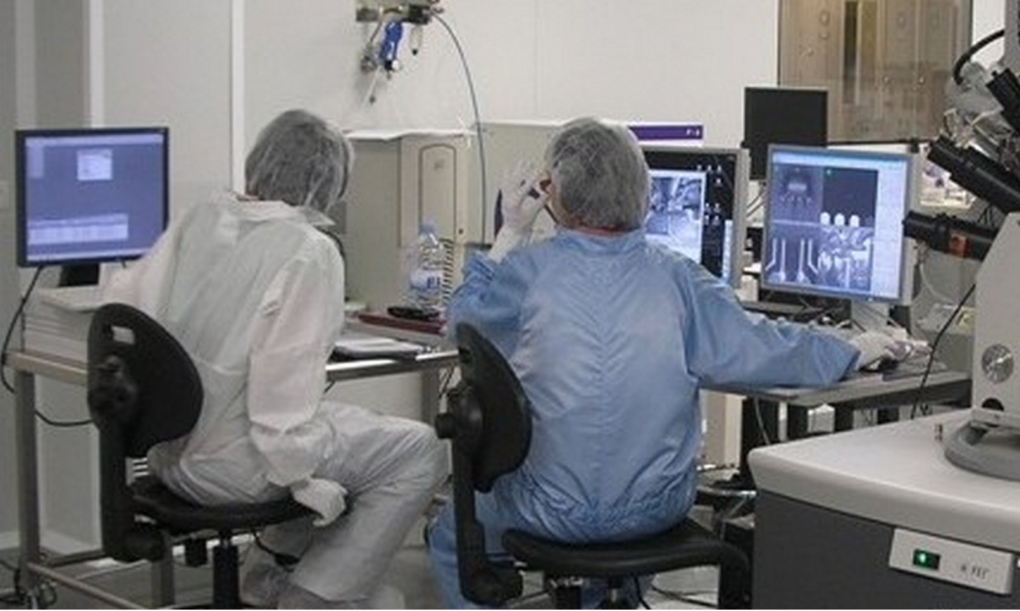
Main Expertise in Nanomagnetism Group is about the Physics of low-dimensional structures, nanostructures, and nanoscale structured complex systems in which nanomagnetism and magnetic characterization, especially magneto-optical methods are the main topics. Main Infrastructures: Clean Room of 300 m2, EBL Raith-150-TWO/E-Line, MOKE microscope, an implementing D-MOKE analysis.
The Nanomagnetism Group is conducting basic and applied world-class research in the field of magnetism in nanoscale structures. The group staff has long-standing expertise and a proven track record in fundamental and applied aspects of nanomagnetism, magnetic materials, and magnetic characterization, especially magneto-optical methods.
Principal investigator:

Dr Paolo Vavassori
IKERBASQUE Research Professor – co-head of the Nanomagnetism Group at CIC nanoGUNE BRTA
Expertise: Magnetic properties characterization, Ferromagnetic resonance, Magnetic force microscopy (MFM).
p.vavassori(at)nanogune.eu
Equipment:
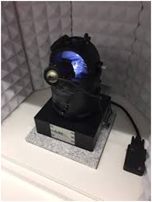
MOKE microscope
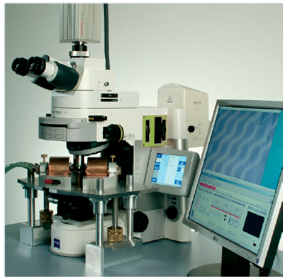
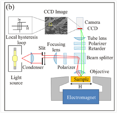
Optical wide-field polarization microscope optimized for Kerr microscopy (Evico Magnetics GmbH, Germany). The microscope is equipped with electromagnets that allow the application of a magnetic field of up to 500 mT (100 mT) along an arbitrary direction in the sample plane (perpendicular to the sample plane) and with a high sensitivity CCD camera that is capable of taking magnetic-contrast images of the sample surface with a spatial resolution of the order of 500 nm. The key feature of our MOKE microscope is that we can measure the field-dependent local magnetization, i.e., a local hysteresis loop, by selecting an arbitrary (shape, size, and position in the field of view) region of interest (ROI), by means of a reduced number of pixels within the CCD camera array, and use it as a light intensity detector for conventional magnetometry [M(H) looper]. The system is equipped with a cryostat (80K – 350 K) and a heater (300 K – 800 K) for temperature-dependent magnetic characterization. Samples can be easily contacted for magneto-transport measurements using the available Keithley 2611A SourceMeter (V range 200 mV – 200 V, I range 100 nA – 10 A, V resolution 5 mV – 5 mV, I resolution 2 pA – 200 mA). Sample size (maximum) 1.0 x 1.0 cm2 (0.7 x 0.7 cm2.for temperature-dependent measurements).
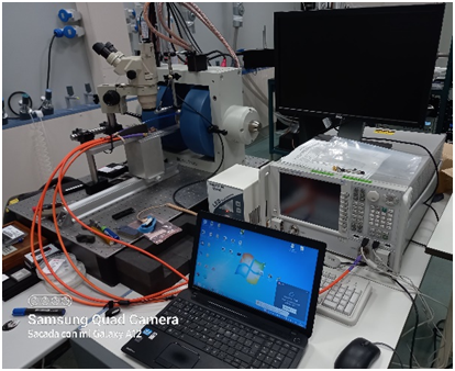
AFM/MFM/HD-KFM Microscopes
Photos of two nanoGUNE AFM/MFM/KFM tools (3 tools in total).
Tools (3) for atomic and magnetic force microscopy (AFM/MFM), and High-Definition Kelvin Force Microscopy (HD-KFM). Different sample holders and probes are available to carry out measurements varying the temperature of the sample (300 K – 650 K using an environmental chamber) and applying voltages to the sample and/or the probe. One tool is equipped with an ultra-sensitive implementation of Kelvin force microscopy, HD-KFM, to measure the surface potential between an oscillating conductive tip and the surface. The surface potential measurement can be used to determine intrinsic properties such as the working function or the band gap and to achieve high resolution and ultra-sensitive MFM images. Sample size (maximum) 1.5 x 1.5 cm2.
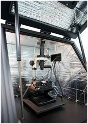
VNA-FMR
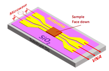
Vector network analyzer-based FMR spectrometer. The sample is placed face-down on a 50 Ω coplanar waveguide. Magnetic-field-dependent perturbation of the transmitted microwave signal is measured over a frequency range of 0.5-40 GHz. The maximum magnetic field (in-plane or out of plane) is 1T. Data are fitted to the Landau-Lifshitz equation. The high sensitivity allows for the detection of localized magnons in nano/microstructures samples. Sample size (maximum) 0.7 x 0.7 cm2.